Advertisement
Grab your lab coat. Let's get started
Welcome!
Welcome!
Create an account below to get 6 C&EN articles per month, receive newsletters and more - all free.
It seems this is your first time logging in online. Please enter the following information to continue.
As an ACS member you automatically get access to this site. All we need is few more details to create your reading experience.
Not you? Sign in with a different account.
Not you? Sign in with a different account.
ERROR 1
ERROR 1
ERROR 2
ERROR 2
ERROR 2
ERROR 2
ERROR 2
Password and Confirm password must match.
If you have an ACS member number, please enter it here so we can link this account to your membership. (optional)
ERROR 2
ACS values your privacy. By submitting your information, you are gaining access to C&EN and subscribing to our weekly newsletter. We use the information you provide to make your reading experience better, and we will never sell your data to third party members.
Analytical Chemistry
Liquid Biopsies Are On The Way
Measurements of cell-free DNA in body fluids could replace tissue biopsies for some applications
by Celia Henry Arnaud
July 7, 2014
| A version of this story appeared in
Volume 92, Issue 27

Cancer biopsies are invasive and often painful procedures in which tissue is removed to diagnose and characterize cancer. And after all the trouble involved in taking biopsies, some still don’t provide physicians with enough tumor cells for further molecular analysis. But a day might be coming when biopsies won’t automatically mean removal of tissue. Instead, they will be performed with samples of easily obtained body fluids such as blood or urine. Advances in DNA testing technology are hastening the arrival of such liquid biopsies.
TRANSPLANT MEDICINE
Liquid Biopsies With Circulating Cell-Free DNA Detect Acute Rejection In Heart Transplants
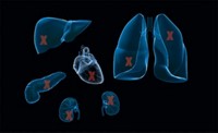
Acute rejection occurs in the first two years for about one-quarter to one-third of heart transplants. If rejection is severe enough, it can result in loss of a transplanted organ.
The most common way to test for rejection is to thread a biopsy instrument through the neck vein and remove tissue for histological analysis. “That procedure has inherent risks and complications,” says Kiran K. Khush, a cardiologist at Stanford University School of Medicine. “It’s uncomfortable for the patient, expensive, and time-consuming.” To top it off, biopsy isn’t even a particularly good gold standard. “You’re sampling such a small piece of tissue that there’s potential to miss rejection when it’s present,” she says.
There is thus a need for a better way to diagnose rejection in time to treat it. Khush and her coworkers recently reported their use of circulating cell-free DNA to diagnose heart transplant rejection.
When cells die, they release their contents into circulation. There’s a baseline level of cell contents in blood as a result of normal cell turnover. In organ transplants, the donor’s DNA differs from the patient’s, and increased levels of donor DNA can be a sign of rejection.
In a three-year study of adult and pediatric heart transplants, Khush and her colleagues demonstrated that the amount of cell-free donor DNA in patients’ plasma can be identified and accurately quantified (Sci. Transl. Med. 2014, DOI: 10.1126/scitranslmed.3007803).
“We show that donor-specific cell-free DNA increases in the setting of acute rejection,” Khush says. “It starts going up several weeks to months before rejection is diagnosed on biopsy.”
To do the analysis, they take blood samples from both the donor and the recipient at the time of transplantation. They sequence highly variable DNA regions to find differences that can later be used to distinguish between donor and patient DNA.
“We found that our cell-free DNA assay was very accurate in picking up moderate to severe rejection,” Khush says. Mild rejection usually resolves on its own without treatment. Higher levels usually require extra immunosuppression therapy.
The assay still needs further clinical trials before it will be ready for regular use on its own. But Khush notes that it should be applicable to any solid-organ transplant. “We were specifically looking at heart transplantation, but the same type of assay should be relevant to kidney, liver, and lung transplantation,” Khush says.
Current research on liquid biopsies for oncology is focused on using techniques such as polymerase chain reaction (PCR) measurements of cell-free DNA to guide and monitor treatment. At this point, scientists doubt the technology will be suitable for initial cancer screening, but they hope it will be able to confirm a diagnosis or help when tumor tissue isn’t available.
When cells die, they release DNA into the blood. There’s always a baseline level of cell-free DNA as a result of normal cell turnover. But disease processes shed disease-specific DNA into circulation, which can be collected and analyzed. Such approaches are already used in prenatal testing because fetal DNA can be found in the mother’s blood, and early work shows promise for improved monitoring of cancer treatment as well.
A key PCR technology being studied for analysis of cell-free DNA is called droplet-based digital PCR. In this approach, a sample is partitioned into thousands or even millions of droplets within an emulsion that also contains the reagents needed for PCR amplification and fluorescent labeling of target DNA.
Droplet-based digital PCR technologies developed by Bio-Rad Laboratories, in Hercules, Calif., and RainDance Technologies, in Billerica, Mass., use nanoliter- and picoliter-scale droplets, respectively. In the case of cancer mutations, mutant DNA is fluorescently labeled with one color and normal DNA is labeled with another. The droplets are detected and counted by an instrument similar to a flow cytometer. The number of droplets with a particular fluorescent label can then be used to calculate the concentration of the target sequence.
“The advantage of using droplets is in the flexibility and the scalability that these droplets enable,” says Viresh Patel, senior marketing manager at Bio-Rad’s Digital Biology Center. “You can scale up or down the number of droplets as the application requires or as needs change.”
Earlier attempts at developing less invasive biopsies involved collecting circulating tumor cells (CTCs). Oncologists have shown that they can use CTCs, which are thought to have broken off solid tumors, as a prognostic indicator in breast and prostate cancers. But widespread use of CTCs has been hindered by their low concentration in blood and by the lack of scientific consensus of what even constitutes a CTC.
“There’s more cell-free DNA available in a fixed volume of blood than there would be CTCs,” Patel says. “In 1 mL of blood there can be up to 10,000 genome equivalents of cell-free DNA.” The same volume of blood is likely to have fewer than 10 CTCs.
Another frustration with CTCs is their delicacy, says Geoffrey R. Oxnard, an oncologist at Dana-Farber Cancer Institute in Boston. “They are hard to handle,” he says. “We want an assay that uses a stable, easy-to-handle molecule. That’s DNA, which is the ultimate stable molecule.” On top of that, the best biomarkers in oncology denote genotypes, genes that encode specific functions, making DNA sequence the perfect analytical target, he adds.
Oxnard, translational research head Cloud P. Paweletz of Dana-Farber’s Belfer Institute for Applied Cancer Science, and coworkers are studying whether mutations detected in cell-free DNA can be used to monitor treatment of lung cancer patients.
“I look for specific oncogenic mutations that I know have clinical meaning,” Oxnard says. In particular, he looks for mutations in the egfr,braf, or kras genes, all of which have implications for lung cancer treatment.
The goal is to be able to replace invasive biopsies for treatment monitoring. PCR-based blood tests for target DNA are both noninvasive and relatively quick, attributes that give them the potential to be better than invasive tumor genotyping. Oxnard and coworkers hope to prove those advantages in validation studies.
In one recent study of lung cancer patients with egfr mutations, they saw changes in the levels of mutations and corresponding cancer progression earlier than they could observe them with imaging methods, in some cases as much as 16 weeks earlier (Clin. Cancer Res. 2014, DOI: 10.1158/1078-0432.ccr-13-2482). But they have not yet used the droplet-based digital PCR assays alone. “We haven’t as part of this clinical research skipped any biopsies or skipped any standard of care,” Oxnard says.
Liquid biopsies are being investigated for other types of cancer as well. David Polsky, a dermatologist at New York University’s Langone Medical Center, is investigating whether droplet-based digital PCR of braf and nras mutations in cell-free DNA can be used as biomarkers of treatment response in patients with metastatic stage IV melanoma.
In research he reported at the American Society of Clinical Oncology last month, Polsky found that droplet-based digital PCR was able to measure clinically relevant changes in levels of braf and nras mutations three months ahead of imaging tests.
Polsky also compared the droplet-based digital PCR assay with the assay for lactate dehydrogenase, an enzyme that is not specific to cancer but is nonetheless used to help determine the stage of cancer. However, LDH is known to be relatively insensitive to changes in disease, Polsky says. “We found that the dynamic range of the cell-free DNA reading was much greater than the dynamic range of LDH,” he says. “You could see that the cell-free DNA was more responsive to changes in the patient’s disease burden.”
Blood is not the only fluid that can be used for liquid biopsies. Trovagene, a molecular diagnostics company in San Diego, focuses on the analysis of cell-free DNA in urine.
“Looked at abstractly, urine is cell-free blood—plasma,” says Antonius Schuh, Trovagene’s chief executive officer. The collection of urine “is entirely noninvasive, and you can get very large samples. Whenever you’re after very rare events, a large sample that you can get often is a good idea.”
Trovagene’s cancer diagnostic tests use droplet-based digital PCR. Depending on whether they’re targeting one or multiple mutations, they detect target DNA with a fluorescent label or by next-generation sequencing.
One advantage of urine relative to blood for cell-free DNA analysis is that DNA can reach higher concentrations in urine. In plasma, the half-life of cell-free DNA is about an hour. The equilibrium signal is therefore likely to be weak. In urine, the signal accumulates over time, potentially making it more easily detectable.
Unlike blood analysis, which reports on DNA changes in the past hour only, measurements of DNA in urine report on longer time periods of up to eight or 10 hours, Schuh says.
The cell-free DNA in urine consists of smaller fragments than are found in blood. That required Trovagene to develop an enrichment assay for DNA fragments with lengths as short as 30 bases.
Trovagene has demonstrated its technology in tests for Erdheim-Chester and Langerhans diseases, rare cancerlike conditions characterized by the proliferation of histiocytes and macrophages, types of white blood cells. Both of these diseases are associated with braf mutations.
“In a data set with 26 patients, we significantly outperformed the biopsy when it came to determining the oncogene mutational status of the patients,” Schuh says. Trovagene offers its diagnostic test through its accredited clinical lab.
Schuh notes that developments in liquid biopsies are being driven by oncologists. “It’s not the companies; it’s not policymakers,” he says. “It’s primarily the oncologists, who see patients every day, who are asking why do we have to fly blind while the surgeons have high-resolution imaging?”


Join the conversation
Contact the reporter
Submit a Letter to the Editor for publication
Engage with us on Twitter