Advertisement
Grab your lab coat. Let's get started
Welcome!
Welcome!
Create an account below to get 6 C&EN articles per month, receive newsletters and more - all free.
It seems this is your first time logging in online. Please enter the following information to continue.
As an ACS member you automatically get access to this site. All we need is few more details to create your reading experience.
Not you? Sign in with a different account.
Not you? Sign in with a different account.
ERROR 1
ERROR 1
ERROR 2
ERROR 2
ERROR 2
ERROR 2
ERROR 2
Password and Confirm password must match.
If you have an ACS member number, please enter it here so we can link this account to your membership. (optional)
ERROR 2
ACS values your privacy. By submitting your information, you are gaining access to C&EN and subscribing to our weekly newsletter. We use the information you provide to make your reading experience better, and we will never sell your data to third party members.
Analytical Chemistry
Imaging Method Captures Structural Details Of Nanoparticles In Liquids
Materials: Researchers use electron microscopy to generate 3-D renderings of freely floating particles
by Mitch Jacoby
July 16, 2015
| A version of this story appeared in
Volume 93, Issue 29

\

\
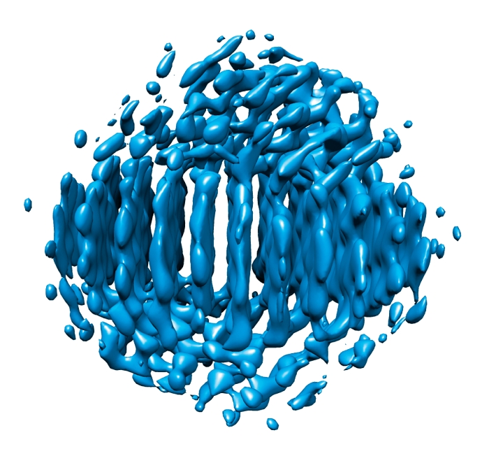
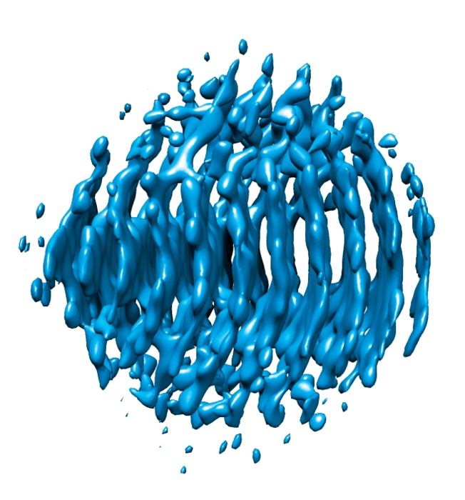
The riblike structures seen here depict atomic planes in two nonidentical platinum particles (2 nm in diameter), which were imaged while they drifted and rotated freely in solution.
Fidgety kids and skittish birds don’t sit still long enough to have their pictures taken. Neither do nanoparticles floating and rotating around in solution. But that hasn’t stopped a team of researchers from producing three-dimensional images of angstrom-sized metal particles in solution at near-atomic resolution (Science 2015, DOI: 10.1126/science.aab1343).
The imaging technique used by the researchers, based on transmission electron microscopy (TEM), may enable scientists to monitor dynamics of individual particles within a colloid in their native state. Structural details gleaned from the imaging method may also lead to new uses for nanoparticles in catalysis, biological imaging, and other areas.
TEM generates 2-D projections of 3-D objects. One way microscopists produce 3-D TEM images is by painstakingly piecing together multiple 2-D images of a rigidly supported sample as it is tilted at various angles. Another way is to record images of many identical particles trapped in various orientations in ice. But those methods aren’t suitable for high-resolution imaging of nanoparticles that are freely rotating in solution.
So A. Paul Alivisatos and Alex Zettl of the University of California, Berkeley; Jungwon Park and David A. Weitz of Harvard University; and coworkers devised a way to sidestep that limitation.
Drawing on a liquid-sampling technique the researchers reported in 2012 , they packaged a few droplets of colloidal platinum nanoparticles in a nanosized graphene bubble. Then they used a microscope equipped with a highly sensitive detector to zoom in on individual particles and record many short-exposure images.
Because the particles were constantly moving, the images were inherently low quality. But by applying a computational technique the researchers developed for this purpose, they were able to produce high-resolution 3-D reconstructions of randomly moving nonidentical particles.
This work is likely to make an impact in biological imaging, catalysis, and other fields, says Joerg R. Jinschek, a TEM specialist for microscope maker FEI who is based in the Netherlands. Jinschek explains that attaching a colloidal particle to a rigid surface—as has been done for imaging purposes in the past—can alter its structure. Because structure and other properties often depend on a particle’s state or environment, researchers want detailed information about nanoparticles in their natural states, Jinschek says. He adds that being able to image freely moving nanoparticles in liquids, as was done in the new study, will help scientists determine relationships between a nanoparticle’s structure and its function.


Join the conversation
Contact the reporter
Submit a Letter to the Editor for publication
Engage with us on Twitter