Advertisement
Grab your lab coat. Let's get started
Welcome!
Welcome!
Create an account below to get 6 C&EN articles per month, receive newsletters and more - all free.
It seems this is your first time logging in online. Please enter the following information to continue.
As an ACS member you automatically get access to this site. All we need is few more details to create your reading experience.
Not you? Sign in with a different account.
Not you? Sign in with a different account.
ERROR 1
ERROR 1
ERROR 2
ERROR 2
ERROR 2
ERROR 2
ERROR 2
Password and Confirm password must match.
If you have an ACS member number, please enter it here so we can link this account to your membership. (optional)
ERROR 2
ACS values your privacy. By submitting your information, you are gaining access to C&EN and subscribing to our weekly newsletter. We use the information you provide to make your reading experience better, and we will never sell your data to third party members.
Environment
Sorting neurons from preserved brain tissue
New methods allow researchers to sort cells in chemically fixed, frozen brain tissue by using flow cytometry
by Wudan Yan
April 3, 2017
| A version of this story appeared in
Volume 95, Issue 14
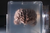
Neuroscientists routinely store brain tissue samples in brain banks for future analysis. Being able to use cell-sorting techniques such as flow cytometry on this preserved tissue would allow researchers to study the effects of drugs, environmental factors, and diseases on specific brain cell types. However, it’s difficult to separate cells from chemically fixed, frozen tissue samples and prepare them for flow cytometry without causing damage. Now, Charles D. Nichols of the Louisiana State University Health Sciences Center New Orleans and colleagues have developed new protocols to overcome these limitations (ACSChem. Neurosci. 2017, DOI: 10.1021/acschemneuro.6b00374). The researchers separated cells from human and rodent brain tissue samples—fixed using preservatives such as formalin, zinc, and paraformaldehyde—by passing liquefied frozen tissue through needles multiple times and applying an enzyme solution that cleaves chemical bonds between cells. They then fluorescently tagged the cells and passed cell suspensions through a flow cytometer to isolate two types of neurons. Importantly, the researchers were able to perform downstream genetic and biochemical analyses on these recovered cells, indicating the cells were undamaged.


Join the conversation
Contact the reporter
Submit a Letter to the Editor for publication
Engage with us on Twitter