Advertisement
Grab your lab coat. Let's get started
Welcome!
Welcome!
Create an account below to get 6 C&EN articles per month, receive newsletters and more - all free.
It seems this is your first time logging in online. Please enter the following information to continue.
As an ACS member you automatically get access to this site. All we need is few more details to create your reading experience.
Not you? Sign in with a different account.
Not you? Sign in with a different account.
ERROR 1
ERROR 1
ERROR 2
ERROR 2
ERROR 2
ERROR 2
ERROR 2
Password and Confirm password must match.
If you have an ACS member number, please enter it here so we can link this account to your membership. (optional)
ERROR 2
ACS values your privacy. By submitting your information, you are gaining access to C&EN and subscribing to our weekly newsletter. We use the information you provide to make your reading experience better, and we will never sell your data to third party members.
Biological Chemistry
A new view of the spliceosome
Structural biologists capture important state of cellular machine responsible for making humans more complex than worms
by Sarah Everts
May 24, 2017
| A version of this story appeared in
Volume 95, Issue 22

Humans share a comparable number of protein-coding genes with the simple roundworm Caenorhabditis elegans, yet we are arguably more sophisticated organisms. This difference in complexity is thanks to the spliceosome, an enormous piece of biochemical machinery found in the nucleus.
The spliceosome cuts out unnecessary sequences of freshly transcribed RNA called introns and joins together the remaining sections to form messenger RNA (mRNA). The variable way that the spliceosome combines the nonintron RNA results in about ten times as many proteins in human cells as the number of genes in our genome.
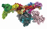
Now, thanks to cryo-electron microscopy, researchers have a new, near-atomic-level view of this cellular machine in its precatalytic state, before it has made the decision to start splicing RNA (Nature, 2017, DOI: 10.1038/nature22799).
Dozens of proteins and five protein-RNA complexes, called ribonucleoproteins, come and go as the spliceosome prunes RNA after it has been transcribed from DNA and before protein production begins.
During splicing, there are seven colossal rearrangements of the spliceosome during its assembly, activation, and catalysis. The current research—performed by Clemens Plaschka, Pei-Chun Lin, and Kiyoshi Nagai at the Medical Research Council Laboratory of Molecular Biology—captured the spliceosome in the particularly important first arrangement when the machinery has loaded unspliced RNA but hasn’t yet gotten down to catalysis, comments Yigong Shi at Tsinghua University, who wasn’t involved in the work. This is a commitment step, an important decision-making point in splicing.
The team found that 24 proteins associated with the spliceosome help keep the machine in a precatalytic state. Thereafter, these 24 proteins depart and another 22 arrive to help the spliceosome machinery undergo an extensive rearrangement in preparation for catalysis, Plaschka says. The work provides “a framework to dissect the activation mechanism and to determine the precise order of molecular events leading to formation of the spliceosome active site,” the researchers write in the paper.
To date, four subsequent spliceosome states have been captured by structural biologists at near atomic resolution, Shi adds. “This fifth structure is an important step closer to recapitulation of the entire catalytic splicing cycle.”
This article has been translated into Spanish by Divulgame.org and can be found here.


Join the conversation
Contact the reporter
Submit a Letter to the Editor for publication
Engage with us on Twitter