Advertisement
Grab your lab coat. Let's get started
Welcome!
Welcome!
Create an account below to get 6 C&EN articles per month, receive newsletters and more - all free.
It seems this is your first time logging in online. Please enter the following information to continue.
As an ACS member you automatically get access to this site. All we need is few more details to create your reading experience.
Not you? Sign in with a different account.
Not you? Sign in with a different account.
ERROR 1
ERROR 1
ERROR 2
ERROR 2
ERROR 2
ERROR 2
ERROR 2
Password and Confirm password must match.
If you have an ACS member number, please enter it here so we can link this account to your membership. (optional)
ERROR 2
ACS values your privacy. By submitting your information, you are gaining access to C&EN and subscribing to our weekly newsletter. We use the information you provide to make your reading experience better, and we will never sell your data to third party members.
Cancer
Chemistry In Pictures
Chemistry in Pictures: Milky Way malady
by Alexandra Taylor
April 7, 2020

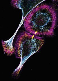
This image, taken with a confocal microscope, shows a pancreatic cancer cell. The cell’s irregular surface is covered with adhesions, tiny structures that help the cell attach to other cells and interact with its environment. The long, thin strands are microtubules that make up the cell’s cytoskeleton. Lorna Young, a postdoc with the Institute of Translational Medicine at the University of Liverpool, captured this image. Young’s team studies how healthy and diseased cells move within the body.
Submitted by Lorna Young
Do science. Take pictures. Win money. Enter our photo contest here.


Join the conversation
Contact the reporter
Submit a Letter to the Editor for publication
Engage with us on Twitter