Advertisement
Grab your lab coat. Let's get started
Welcome!
Welcome!
Create an account below to get 6 C&EN articles per month, receive newsletters and more - all free.
It seems this is your first time logging in online. Please enter the following information to continue.
As an ACS member you automatically get access to this site. All we need is few more details to create your reading experience.
Not you? Sign in with a different account.
Not you? Sign in with a different account.
ERROR 1
ERROR 1
ERROR 2
ERROR 2
ERROR 2
ERROR 2
ERROR 2
Password and Confirm password must match.
If you have an ACS member number, please enter it here so we can link this account to your membership. (optional)
ERROR 2
ACS values your privacy. By submitting your information, you are gaining access to C&EN and subscribing to our weekly newsletter. We use the information you provide to make your reading experience better, and we will never sell your data to third party members.
Cancer
Chemistry In Pictures
Chemistry in Pictures: The ‘bones’ inside your bones
by Manny I. Fox Morone
February 25, 2021

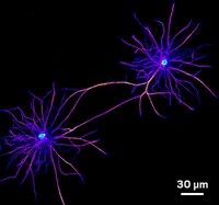
The inner workings of these bone cells as they divide appear striking under the microscope, but that’s not why Lorna Young wants a closer look at them. Young, a postdoc in Tobias Zech’s lab at the University of Liverpool, studies how the cytoskeleton coordinates cellular behavior in various diseases like in osteosarcoma, a bone cancer that’s afflicting these cells. Using fluorescent small molecules and primary antibodies, she’s able to focus on a few parts of cytoskeletal structure that she likes to refer to as the "bones" of cells, including actin (magenta), microtubules (yellow), intermediate filaments (cyan), under 40× magnification.
Submitted by Lorna Young/Tobias Zech lab. Follow Lorna on Twitter @drlornayoung
Do science. Take pictures. Win money. Enter our photo contest here.


Join the conversation
Contact the reporter
Submit a Letter to the Editor for publication
Engage with us on Twitter