Advertisement
Grab your lab coat. Let's get started
Welcome!
Welcome!
Create an account below to get 6 C&EN articles per month, receive newsletters and more - all free.
It seems this is your first time logging in online. Please enter the following information to continue.
As an ACS member you automatically get access to this site. All we need is few more details to create your reading experience.
Not you? Sign in with a different account.
Not you? Sign in with a different account.
ERROR 1
ERROR 1
ERROR 2
ERROR 2
ERROR 2
ERROR 2
ERROR 2
Password and Confirm password must match.
If you have an ACS member number, please enter it here so we can link this account to your membership. (optional)
ERROR 2
ACS values your privacy. By submitting your information, you are gaining access to C&EN and subscribing to our weekly newsletter. We use the information you provide to make your reading experience better, and we will never sell your data to third party members.
Analytical Chemistry
Assaying Antibodies
Drug manufacturers pinpoint techniques to analyze growing and diverse class of therapeutics
by Jyllian Kemsley
January 16, 2012
| A version of this story appeared in
Volume 90, Issue 3

Monoclonal-antibody-based drugs are a large and growing segment of pharmaceuticals. Since muromonab-CD3 (Orthoclone OKT3) was first approved by the Food & Drug Administration in 1986 as an antirejection drug for organ transplant patients, more than 30 antibody-based drugs have entered the market, and many more are in the pipeline. In 2011 alone, FDA approved belimumab (Benlysta) for lupus, ipilimumab (Yervoy) for metastatic melanoma, and brentuximab vedotin (Adcetris) for two types of lymphoma. Market research company Datamonitor estimates that sales of antibody therapeutics will grow by 8.2% from 2010 to 2016, the fastest of any therapeutic class, with sales expected to surpass $65 billion by 2016.
Although drugs based on monoclonal antibodies—MAbs—have widely varied therapeutic action, they are all based on the same protein, immunoglobulin G (IgG). But this shared attribute does not mean that these drugs are simple to assay. MAbs are composed of more than 1,000 amino acids in four peptide chains, and they bear sugars and other chemical modifications.
As the MAb class grows, scientists are settling on some standard critical protein qualities and analytical testing approaches for drug identity, quality, and purity for these large, complex molecules.
Along those lines, the U.S. Pharmacopeial Convention (USP), the pharmaceutical standard-setting organization in the U.S., is in the midst of developing “best practice” guidelines and regulatory standards for MAb drug developers. “The industry has more than 20 years of manufacturing experience with these types of molecules, and we’re seeing products that are extremely pure and really well characterized,” says Tina S. Morris, USP’s vice president for biologics and biotechnology. “Every antibody has slightly different purification and analytical characteristics, but you’re never starting from zero with a completely unknown protein.”
One set of standards, to be known as Chapter 1260, will focus on how to develop, define quality attributes for, and manufacture MAbs.
These standards will not be legally binding on MAb manufacturers but instead are intended to provide worldwide best practice guidance, says Anthony Mire-Sluis, corporate vice president for product and device quality at Amgen and chair of the USP Recombinant Therapeutic Monoclonal Antibodies Expert Panel, which is developing the standards. The expert panel includes 13 industry members, as well as seven representatives from regulatory and other standard-setting agencies.
The other set of standards, Chapter 129, will be legally binding on MAb manufacturers and outlines the minimal quality attributes common to all antibodies and the corresponding recommended analytical tests for MAb therapeutics. Several companies will validate the proposed methods to ensure that they work, and the standards will also include limits derived from manufacturer data on actual products. But, Mire-Sluis notes, companies will be able to justify using different tests or limits as part of their drug application package if they think their product merits something atypical.

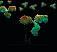
Structurally, an antibody, or immunoglobulin, is a large, Y-shaped protein composed of two “heavy” and two “light” peptide chains. The heavy chains come together at the bottom half of the Y, also called the crystallizable fragment, or Fc. Disulfide bonds and noncovalent interactions hold the chains together. This part of the antibody has a conserved amino acid sequence and binds to receptors to activate parts of the immune system. Different antibody classes—IgA, IgD, IgE, IgG, and IgM—have different heavy chains that determine specific immunological roles. IgG provides most of the body’s immunity to invading pathogens.
At the top half of the antibody Y, the heavy chains branch out and each associates with a light chain, also through disulfide bonds and noncovalent interactions. This region of the antibody is called the antigen-binding fragment, or Fab, and it is here that the amino acid sequence varies to bind to assorted foreign molecules: Both arms of a specific antibody have the same amino acid sequence, while the variable portion differs between different antibody molecules.
But because MAbs are made by cloned cell lines with identical cells, all of the antibodies produced by a cell line engineered for producing a particular therapeutic should be identical. Monoclonal IgG therapeutics were originally based on mouse antibodies, but they now can be chimeric, which means a part-mouse/part-human sequence, or “fully” human. Structurally, the three types are very similar, but they may provoke different immune responses in patients.
In addition to their basic structural likeness, MAb therapeutics also tend to be purified similarly, USP’s Morris notes. Purification typically starts with affinity chromatography using a solid phase that anchors protein A, a bacterial protein that selectively binds to the antibodies’ conserved Fc region. After that, the drug is likely treated to inactivate any contaminating viruses and may undergo fine-tuning purification steps. Most antibodies are also currently produced from two popular cell lines, which means that their impurity profiles are similar. The combination of similar cell culture and purification, on top of targeting the same protein, further lends analysis of MAb therapeutics to similar protocols, Morris says.
But despite these similarities, MAbs may show heterogeneity depending on how they were produced and processed by their originating cells. Problems with DNA translation or transcription may lead to different protein sequences. Proteins may misfold or mismatch their disulfide bonds. Posttranslational modification may produce different glycosylation patterns. Side chains may also be subject to chemical reactions, such as oxidation or deamidation. Proteolytic enzymes may clip the protein, especially the lysines at the ends of the heavy chains in the Fc. MAbs may also denature or aggregate as they go through purification or formulation.
Because of these effects, even a perfectly prepared MAb therapeutic will include IgG proteins with varied structures and properties, says William Whitford, senior market manager for cell culture and bioprocessing at Thermo Fisher Scientific. “Unlike small-molecule drugs, biologics show a distribution of slightly different molecules,” he says. The analytical challenge is therefore to ensure that the population distribution doesn’t vary significantly from the drug that was developed, tested clinically, and approved.
Some properties are a process red flag. Oxidation or deamidation, for example, by itself generally doesn’t affect an antibody’s clinical function, says Raymond Kaiser, global science leader and vice president for biotechnology services at Covance, a contract drug development company. But variability in those categories is usually a sign that the manufacturing process is not well controlled, and other protein characteristics, such as aggregation, may be affected, Kaiser says.
Other modifications are a clinical concern. A misfolded protein or one with abnormal disulfide bonds probably won’t have the structure needed to function properly. Additionally, IgGs have a conserved glycosylation site on the Fc, but variations in those sugars and glycosylation elsewhere on the protein may elicit an unwanted immunological response, such as anaphylaxis. Protein aggregates may also provoke an immune response.
And so a suite of analytical tests is necessary to ensure that a MAb therapeutic is within the bounds of the drug that was originally prepared, tested, and approved. “You live and die by your analyticals,” Kaiser says. “If you don’t have your analyticals, you don’t know anything.”
The first order of business is often to identify whether a manufacturer has made the correct protein. One common approach is to do an immunoassay, such as an enzyme-linked immunosorbent assay (ELISA). In these types of assays, a stationary solid phase is loaded with a molecule that will bind specifically to the target antibody. A drug sample is loaded, incubated, and washed, and some sort of label is used to assay whether and how much of the target antibody is bound.
Another option for identifying proteins is peptide mapping, which involves using a protease enzyme to cleave an antibody into characteristic segments that can then be separated by gel electrophoresis or column chromatography to yield a particular fingerprint.
On the quality front, tests such as ion-exchange chromatography, isoelectric focusing, or capillary electrophoresis will pick up variations in charge within the protein structure that might indicate oxidation, deamidation, or protein clipping. In ion-exchange chromatography, how proteins interact with a charged stationary phase determines how quickly they elute. Isoelectric focusing takes advantage of the fact that protein charges are a function of pH and separates proteins using a gel with a pH gradient. Capillary electrophoresis separates proteins by applying an electric field to a capillary tube and can be done using either free solution (capillary zone electrophoresis) or a gel (capillary gel electrophoresis).
For glycosylation, manufacturers often create a glycan map similar to a protein map. The antibody is enzymatically or chemically treated to remove the sugars, which are then given a fluorescent tag and separated using high-performance or ultraperformance liquid chromatography. The glycans’ identities determine when they elute from the column. Sialic acids, however, need a different approach. Because they are destroyed by the standard acid treatment to remove sugars, they are instead removed using mild acid and then analyzed by high-pH anion-exchange chromatography with electrochemical detection.
To further pin down the specifics of chemical modifications or changes in glycosylation patterns, liquid chromatography-mass spectrometry (LC/MS) has become the standard approach. An antibody or its sugars are digested and separated as in other tests, but the resulting fragments are analyzed using mass spectrometry. LC/MS provides exact mass identification of the fragments but requires a highly skilled operator, so it’s mostly used in development or problem solving, says Taegen Clary, marketing manager for biopharmaceuticals at Agilent.
Nevertheless, LC/MS data have become a necessary part of drug application packages to demonstrate that manufacturers understand what they’re seeing in their quality-control suite of assays. “I believe that if you don’t supply some LC/MS data in a package, you’ll be asked for it because it is so common now,” Covance’s Kaiser says.
To pick up aggregation, one approach is simply to look at a protein solution. If the aggregates are big enough, the solution will appear hazy rather than clear. To check for smaller particles, size-exclusion chromatography, also known as gel-filtration chromatography, is the standard method, with detection by either ultraviolet light absorption or multiangle laser light scattering.
Other techniques to look for aggregation include light obscuration and microflow imaging. Microflow imaging is a microscopy technique that allows scientists to distinguish, for example, whether a particle is composed of protein or of silicon oil, which is used in drug formulations as a lubricant for syringes. Those techniques as well as analytical ultracentrifugation might be used in development to help understand antibody aggregation or in production to diagnose a problem, Kaiser says, but the process workhorse is high-throughput size-exclusion chromatography.
Aside from the quality of the IgG molecule, manufacturers must also keep an eye on the purity of the drug solution. Common impurities that manufacturers watch for include residual DNA or other proteins from the host cells, plus any protein A that might have carried through purification. Quantitative real-time polymerase chain reaction (Q-PCR) methods are used to amplify and quantify DNA, while ELISA targets protein A and host-cell proteins. In early development, manufacturers might use cell-line-specific ELISA kits that contain antibodies to common cellular proteins. In late-stage development, however, companies often develop process-specific assays by taking cells that don’t produce the therapeutic through the purification process and using the output to develop antibodies to whatever proteins remain, Kaiser says.
One additional area of MAb analysis is assays for potency. Potency tests, however, cannot readily be standardized across the class. Different clinical actions by necessity require different potency tests, which are often cell-based assays in which adding the drug product elicits some kind of measurable response.
What’s being looked at is also a consideration, whether it’s IgG proteins by themselves; antibody-drug conjugates, which tether a small-molecule drug to an IgG; or antibody fusion proteins, which typically combine the IgG Fc component with another protein. For antibody-drug conjugates, most assays would be similar to those for unconjugated IgGs, but scientists might pay more attention to a peptide mapping assay to make sure they know where and how successfully the small molecule attached, Kaiser says. For fusion proteins, manufacturers would probably turn to the same analytical tools as for intact IgGs, but they would have to do more development work to optimize assays and understand what the data show.
Advertisement
Many of the analytical methods generally used on MAbs, such as ELISA, charge separation procedures, or size-exclusion chromatography, are time-honored biochemical techniques, says Martina Bielefeld-Sevigny, vice president and general manager of drug discovery and research reagents at PerkinElmer. The push now is to make those methods faster and easier, with better precision, accuracy, reproducibility, and sensitivity. In many cases, that means going to microfluidic, lab-on-a-chip platforms that use smaller amounts of materials, provide faster response times, and enable high-throughput analysis.
“I think that the monoclonal industry is a trailblazer,” USP’s Morris says. Pointing as an example to common use of capillary techniques instead of plain gel electrophoresis, she adds, “I think they use fairly sophisticated analytical techniques compared to plasma or peptide industry people.”
That’s something that consumers can appreciate, for the extra assurance that a drug will work as advertised.


Join the conversation
Contact the reporter
Submit a Letter to the Editor for publication
Engage with us on Twitter