Advertisement
Grab your lab coat. Let's get started
Welcome!
Welcome!
Create an account below to get 6 C&EN articles per month, receive newsletters and more - all free.
It seems this is your first time logging in online. Please enter the following information to continue.
As an ACS member you automatically get access to this site. All we need is few more details to create your reading experience.
Not you? Sign in with a different account.
Not you? Sign in with a different account.
ERROR 1
ERROR 1
ERROR 2
ERROR 2
ERROR 2
ERROR 2
ERROR 2
Password and Confirm password must match.
If you have an ACS member number, please enter it here so we can link this account to your membership. (optional)
ERROR 2
ACS values your privacy. By submitting your information, you are gaining access to C&EN and subscribing to our weekly newsletter. We use the information you provide to make your reading experience better, and we will never sell your data to third party members.
Metal-Organic Frameworks
Ultrasound triggers porous nanoparticles to attack tumors in mice
Metal-organic framework forms particle-embedded porphyrin-zinc complexes that generate reactive oxygen species
by Mark Peplow, special to C&EN
April 25, 2018
| A version of this story appeared in
Volume 96, Issue 18

Metal-organic frameworks (MOFs) are already famed for their ability to store and separate gases, but there is growing interest in their potential medical applications. Chinese researchers have now converted a MOF into nanoparticles that harness the power of ultrasound to kill tumor cells in mice (Adv. Mater. 2018, DOI: 10.1002/adma.201800180).
This general approach to attacking cancer is called sonodynamic cancer therapy, and it relies on compounds that are activated by a targeted burst of ultrasound. The sound waves prompt the compounds, known as sonosensitizers, to generate reactive oxygen species that destroy cancer cells in the immediate vicinity, while avoiding systemic side effects.
The strategy is similar to light-based photodynamic therapy, but ultrasound can penetrate deeper into tissue than light, so the strategy, in principle, could treat more inaccessible tumors. Although the technology has been tested in a handful of patients, the field is still in its infancy. “There are very few groups working on it,” says Nikolitsa Nomikou, who develops sonodynamic therapy agents at University College London and was not involved in the new research.
Among the most widely tested sonosensitizers are porphyrin derivatives, which researchers co-opted from photodynamic therapy. These compounds can produce a lot of reactive oxygen species but usually have poor water solubility and tend to be metabolized quickly by the body. Various inorganic nanoparticle sonosensitizers are more stable, but offer relatively poor yields of reactive oxygen species.
Huiyu Liu at the Beijing University of Chemical Technology and colleagues have now combined some of the key benefits of each class of sonosensitizer into a single material that avoids their main pitfalls.
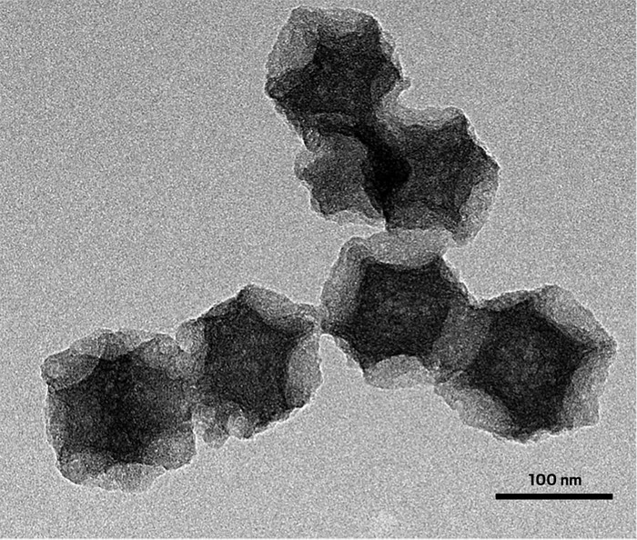
The team’s sonosensitizer is based on a MOF called ZIF-8, which contains a porous lattice of zinc ions held together by imidazolate linkers. After coating ZIF-8 particles with a protective shell of silica to prevent them from sticking together, the researchers heated them at 800 °C for two hours. The MOF transformed into porous carbon nanoparticles that contained zinc coordinated to porphyrin-like rings. Then the researchers stripped away the silica shell and decorated the 140 nm-wide particles with polyethylene glycol to improve their solubility and transport in the body.
A solution of the nanoparticles in water produced large amounts of hydroxyl radicals and singlet oxygen when hit with ultrasound. “The hydroxyl radical generation efficiency is higher than porphyrin-zinc,” says Xueting Pan, a member of the research team.
The precise mechanism involved in generating reactive oxygen species with ultrasound remains one of the big unanswered questions in the field. Previous studies have shown that it involves the formation of tiny bubbles during the ultrasound blast, Pan says. When these bubbles collapse, they can generate flashes of light that excite electrons within a sonosensitizer, ultimately triggering the formation of reactive oxygen species, Nomikou says. The collapsing bubbles can also create jets of fluid that may damage tumor cells, she adds.
The Beijing team found that their nanoparticles’ porous structure helped to seed bubble formation, which may partly explain their success in tests on mice with implanted breast cancer tumors.
Advertisement
After injecting the mice with a solution of the particles, the researchers bombarded the tumor site with 1 MHz ultrasound for five minutes, repeated the ultrasound treatment after 3 days, and then followed the mice’s progress for another 15 days. By the end of that period, the treatment had killed 85% of tumor cells, with no observable side effects or damage to major organs. Ultrasound alone killed just 35% of tumor cells, while injected sonosensitizer particles without any applied ultrasound reduced tumor cell count by just 15%. “It’s very exciting and innovative,” Nomikou says. “It is a system with potential.”


Join the conversation
Contact the reporter
Submit a Letter to the Editor for publication
Engage with us on Twitter