Advertisement
Grab your lab coat. Let's get started
Welcome!
Welcome!
Create an account below to get 6 C&EN articles per month, receive newsletters and more - all free.
It seems this is your first time logging in online. Please enter the following information to continue.
As an ACS member you automatically get access to this site. All we need is few more details to create your reading experience.
Not you? Sign in with a different account.
Not you? Sign in with a different account.
ERROR 1
ERROR 1
ERROR 2
ERROR 2
ERROR 2
ERROR 2
ERROR 2
Password and Confirm password must match.
If you have an ACS member number, please enter it here so we can link this account to your membership. (optional)
ERROR 2
ACS values your privacy. By submitting your information, you are gaining access to C&EN and subscribing to our weekly newsletter. We use the information you provide to make your reading experience better, and we will never sell your data to third party members.
Reaction Dynamics
Ultrafast imaging watches photochemistry frame by frame
Femtosecond X-ray and electron scattering methods reveal a string of never-before-seen molecular events
by Mitch Jacoby
July 19, 2020
| A version of this story appeared in
Volume 98, Issue 28

“In the blink of an eye” may sound fast to most people, but the tenth of a second or so it takes to blink is an eternity to Hyotcherl Ihee. To him, “fast” means a few femtoseconds, or millionths of a billionth of a second—the fleeting time scale of molecular motions.
As a research group leader at Korea Advanced Institute of Science and Technology (KAIST) and South Korea’s Institute for Basic Science, Ihee leads one of a handful of teams of researchers around the world that specialize in tracking the ultrafast dynamics involved in chemical reactions. In recent weeks, these groups have scored big time, publishing several studies that report a string of firsts. The researchers have tracked never-before-seen molecular events, such as the instantaneous electron jump that kicks off light-driven chemical reactions, the coordinated do-si-do danced by electrons and nuclei, and the step-by-step femtosecond motions of atoms bonding to form a trimer.
“The ability to directly follow ultrafast molecular events as they occur on the femtosecond time scale is simply jaw-dropping,” says Michael P. Minitti, a senior staff scientist at SLAC National Accelerator Laboratory, where several seminal ultrafast studies were conducted.
But these studies provide more than a wow factor to those who hear about them and bragging rights to the researchers who run them. One of the main goals of this work is to directly observe in real time the subtle motions of electrons and nuclei as they form molecular bonds or break them. By using techniques that function like stop-action photography, researchers can stitch together snapshots of these dances, frame-by-frame, to construct chemical reaction movies.
The more detailed the movies, the better scientists can understand reaction mechanisms and the elemental steps that guide a reactant to form one or more products, eventually enabling them to steer reactions exclusively toward a desired product.
These are basic science studies, but what scientists learn from them could be applied in many areas. For example, Ihee says, the ability to track transient atomic motions eventually may enable researchers to zoom in on momentary structural changes in proteins and other biomolecules, a key step toward making better pharmaceuticals by understanding how drug molecules dock with their targets.
Minitti adds that subtle details uncovered recently in ultrafast studies of photosynthesis intermediates broaden scientists’ understanding of light absorption, which could help researchers design better solar cells. Similarly, time-resolved studies of inorganic compounds may eventually lead to energy-saving materials that exhibit superconductivity well above the frigid temperatures required by today’s materials.
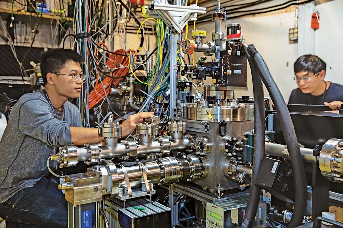
Much of the credit for today’s ultrafast tracking and imaging capabilities goes to advanced instruments, including X-ray free-electron lasers (XFELs) and “electron cameras,” both of which are driven by particle accelerators and housed at large national facilities. The accelerators help generate ultrashort pulses of exceptionally intense X-rays (in XFELs) and electrons (in an electron camera) that are fired at solid, liquid, and gas-phase samples. Both techniques can track ultrafast changes in molecules, giving researchers flexibility to choose the method best suited to the specifics of the investigation.
To record sequences of snapshots, researchers typically use these instruments in a pump-probe arrangement. In that type of experiment, a brief pulse of light, such as from an ultraviolet (UV) laser, “pumps” sample molecules, triggering a rapid change in bonding or other light-induced response.

After firing the pump beam, researchers follow it with a pulse of X-rays or electrons, precisely delayed by some number of femtoseconds. The second shot, which scatters X-rays or electrons from the excited sample, serves as a probe that provides a stop-action photo of the molecular changes set in motion by the pump beam. Then the researchers repeat the one-two punch, systematically increasing the delay between the pump and probe pulses to give a full set of images that capture the molecular process as it unfolds.
That kind of experiment enabled a team of researchers, for the first time, to follow subtle motions of the nuclei in an organic molecule on the femtosecond time scale and subnanometer length scale while simultaneously tracking corresponding changes in the molecule’s electronic structure (Science 2020, DOI: 10.1126/science.abb2235).
“Being able to take those kinds of snapshots is a chemist’s dream,” says Linda Young, a molecular physicist at Argonne National Laboratory and the University of Chicago who was not involved in the study. More importantly, those measurements can help develop theoretical methods to better predict the outcome of chemical reactions, she says.

Young explains that computational chemists have long relied on the Born-Oppenheimer approximation, a nearly 100-year-old simplification for modeling the way molecules behave. It postulates that due to the much lower mass of electrons compared with relatively heavy and sluggish nuclei, the motions of the two types of particles are independent and therefore can be treated separately in calculations. This allows computational scientists to save on computing resources when modeling chemical systems.
But the Born-Oppenheimer approximation often fails in some situations, Young says, such as in photochemistry, electrochemistry, and reactions in which proton and electron transfer are coupled. The new experimental method shows exactly how the motions of the nuclei and electrons are correlated and therefore can boost the accuracy of quantum calculations.
To make the measurements, SLAC’s Jie Yang, Xiaozhe Shen, and Xije Wang, together with Todd J. Martinez of Stanford University and others, zapped pyridine molecules with a UV pump pulse, causing the ringed molecule to undergo rapid puckering motions. Then they probed the excited molecules via ultrafast electron diffraction (UED) by zapping them again, this time with pulses of electrons with millions of electron volts of energy, and recorded how the electrons scattered with SLAC’s electron camera.
UED isn’t new. The original, simpler version of the technique, developed in the 1990s, was pioneered by researchers working with the late Ahmed H. Zewail, a chemistry Nobel laureate. But the earlier experiments missed many of the fine features of the nearly instantaneous structural and electronic changes because the time resolution was only on the order of 10 ps (1 picosecond = 10–12 sec).
By developing an improved version of UED, “we were able to follow the electronic and nuclear changes simultaneously while naturally disentangling the two components,” Martinez says. The “trick” arises from the fact that electrons scatter from molecules, pyridine in this case, in two ways—elastically (without absorbing energy) and inelastically. The elastically scattered beam carries information about nuclear changes. The other beam carries electronic information. The beams emerge from the scattering site at different angles, enabling the researchers to detect them separately and analyze the information they carry.
Other recent studies have taken advantage of brilliant X-ray beams generated by XFELs to reveal never-before-seen light-driven processes. Such phenomena lie at the heart of human vision, photosynthesis, and power generation from solar cells.
In one case, researchers led by Adam Kirrander of the University of Edinburgh and Peter M. Weber of Brown University used an X-ray scattering method to spy on an organic molecule at the moment it absorbed energy from a light pulse—the first step of photoexcitation. The team watched as the molecule’s electron cloud instantaneously ballooned from a relatively confined ground state orbital to a puffy excited state orbital. The event was over in just 30 fs (Nat. Commun. 2020, DOI: 10.1038/s41467-020-15680-4).
The molecule was one the group had studied previously: 1,3-cyclohexadiene (CHD), which is derived from pine oil. Researchers often use CHD to study complex biological reactions, such as the one that produces vitamin D when sunlight shines on skin.
In an earlier study, conducted in 2015, the team pumped the gas-phase molecule with UV light, “which causes CHD to pop open like a zipper,” says SLAC’s Minitti, one of the study’s coauthors. Weber notes that at that time, they were able to track the nuclei over the course of the ring-opening reaction. “But the chemical bonding itself, which is a result of the redistribution of electrons, was invisible. Now the door is open to watching chemical bonds change during reactions,” he says.
“If we want to use light energy to drive chemical transformations, as occurs in natural and artificial photosynthetic systems, we need to understand the earliest stages of light absorption,” says Junko Yano, a molecular biophysicist at Lawrence Berkeley National Laboratory who was not part of the research team. These ultrafast first steps, like the ones analyzed in the recent studies, are important, she explains, because they control the direction and efficiency of the ensuing chemistry.
Ensuing chemistry is exactly what Ihee and coworkers studied at the Pohang Accelerator Laboratory XFEL in South Korea. Ihee’s team prepared a solution of gold cyanide, then used pulses of UV light to stimulate the monomers to form gold-complex trimers [Au(CN)2−]3. That well-known reaction serves as a model for studying photoinitiated processes in solution. As the bond-forming reactions evolved, the team used a scattering method known as X-ray liquidography to scrutinize the process in exquisite detail, focusing on the massive gold atoms. The technique enabled the team to directly and fully track the trajectory of three atoms as they formed a covalently bonded trimer—a feat that had not been accomplished previously (Nature 2020, DOI: 10.1038/s41586-020-2417-3).
The reaction sounds very simple, Ihee says, but it isn’t. Years of time-resolved spectroscopy studies have left key questions unanswered, he says. For example, do the two covalent bonds form simultaneously or sequentially?

Now, thanks to the team’s ultrafast scattering experiment, scientists have the answer—sequentially. Within 60 fs after UV light hits a cluster of three nonbonded monomers—A, B, and C—two of them bond covalently, forming A–B + C. The trio carries on with a number of femtosecond dance steps, and within 360 fs, forms the second bond, resulting in a linear covalently bonded trimer complex A–B–C.
As the large organizations that run XFELs continue to boost these instruments’ capabilities, researchers will likely uncover ever-more subtle molecular phenomena. Although these new findings could someday be applied to fields such as drug discovery, that’s not Ihee’s main goal.
“My research is curiosity-driven rather than goal-driven,” he says. “I believe in the power of basic science to nourish future innovative sciences and technologies.” As an example, he points to magnetic resonance imaging. MRI was not invented on day one as a medical imaging tool, he says. Rather, people were just curious about nuclear spin dynamics. That curiosity led to innovation.
In the short term, curiosity is leading the field of ultrafast dynamics to be able to see “incredibly tiny things on unimaginably short time scales,” Minitti says. “These capabilities are truly game changing. It’s what gets us out of bed in the morning.”
Correction
This story was updated on Aug. 12, 2020, to correct the chemical formula for the gold-complex trimer [Au(CN)2−]3.


Join the conversation
Contact the reporter
Submit a Letter to the Editor for publication
Engage with us on Twitter