Advertisement
Grab your lab coat. Let's get started
Welcome!
Welcome!
Create an account below to get 6 C&EN articles per month, receive newsletters and more - all free.
It seems this is your first time logging in online. Please enter the following information to continue.
As an ACS member you automatically get access to this site. All we need is few more details to create your reading experience.
Not you? Sign in with a different account.
Not you? Sign in with a different account.
ERROR 1
ERROR 1
ERROR 2
ERROR 2
ERROR 2
ERROR 2
ERROR 2
Password and Confirm password must match.
If you have an ACS member number, please enter it here so we can link this account to your membership. (optional)
ERROR 2
ACS values your privacy. By submitting your information, you are gaining access to C&EN and subscribing to our weekly newsletter. We use the information you provide to make your reading experience better, and we will never sell your data to third party members.
Mass Spectrometry
Breaking down biomass with enzymes from an ant farm
Researchers use spatial multiomics to find useful enzymes
by Laurel Oldach
October 23, 2023
| A version of this story appeared in
Volume 101, Issue 35

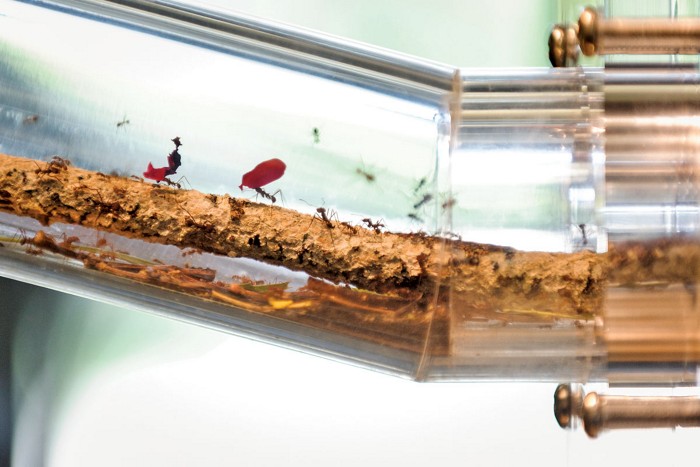
Visitors to Cameron Currie’s laboratory at the University of Wisconsin–Madison used to be mesmerized by a demonstration colony of leaf-cutting ants in the lobby. When the microbiologist announced his plan to move the lab to Ontario, his colleagues asked if the ants had to go too.
Given a handful of oak leaves—which Currie’s lab, now at McMaster University, collects by the garbage bag full—the ants quickly show how they earned their name. With audible crunching, they shred the leaves into tiny pieces and carry them home to their colony. There, the leaf fragments are digested by one of nature’s most efficient systems for breaking down plant cell walls: a fungal garden.
That system turns the steady flow of leaf biomass into nutrition for the ants. Currie and long-term collaborator Kristin Burnum-Johnson, a senior scientist at Pacific Northwest National Laboratory (PNNL), are trying to understand exactly how the garden breaks down hard-to-digest plant polymers. Using a new spatial multiomics method, they can now pinpoint individual enzymatic reactions in the garden. By piecing together how microbial enzymes work for the ants, they hope to gain insights that can be adopted for industrial purposes.
Spongy layers
Many species of ant cultivate fungi, but by far the most elaborate gardeners are Atta texana. To extract energy from the leaves they forage, these ants maintain a crop called Leucoagaricus gongylophorus, a fungus that has never been found anywhere outside the homes of leaf-cutting ants. Researchers estimate that the lives of these two symbionts have been intertwined for 50 million–60 million years, making ant farming older than human agriculture.

“The fungus is kind of [the ants’] house—but also a digestive system,” says Mauricio Caraballo, a researcher in Pieter Dorrestein’s laboratory at the University of California San Diego. Caraballo has worked on mapping the metabolome, the array of small molecules created by digestion in the fungal garden. The garden, which can grow to the size of a basketball, is a spongy structure honeycombed with ant passageways. It forms what Margaret Thairu, a postdoctoral scholar formerly in the Currie lab, calls “a beautiful, stratified structure” at the centerpiece of the ants’ underground nest.
The garden is built as ants bring in leaves foraged from their rainforest surroundings. After cleaning the leaves, the ants deposit them on top of the garden and then add fecal matter rich in digestive enzymes to start the breakdown process. The lower layers of the garden are rife with fungus, which feeds on the leaf matter and grows specialized, nutrient-rich organs called gongylidia. These gongylidia are what the ants feed on.
Finally, leaf fragments and other material that the system has not digested settle to the bottom of the heap, and ants carry them off to a waste repository.
Breaking down lignin
Although it’s clear that the system breaks down leaves, Currie says, for many years the process has been mysterious. Many researchers have focused on understanding the insect-fungus symbiosis, he says, but “the whole process of breaking down plant biomass has been kind of . . . unknown.”
One of the difficulties for researchers has been understanding how the garden microbes crack through plant cell walls—a formidable catabolic challenge. Plants safeguard their cells with an interlaced, covalently cross-linked lattice made mostly of the polymers cellulose, hemicellulose, and lignin. Of the three, lignin is the most robust, says Mariana Barcoto, a graduate student at São Paulo State University (UNESP) who studies the metabolism and microbial ecology of fungal gardens.
Whereas cellulose and hemicellulose are polysaccharides with regular, repeating structures, lignin is complex and amorphous. It is made up of three main aromatic subunits linked irregularly with both C–O and C–C bonds. To make matters more complicated, its subunit stoichiometry differs from one plant to another.
All that heterogeneity, Barcoto says, leads most lignin-decomposing organisms to rely on generalist oxidative mechanisms—including enzymes with wide substrate profiles in the peroxidase, oxidoreductase, and oxidase families. “For breaking cellulose down, you have a very specific set of enzymes. For lignin, you don’t,” Barcoto says.
Lignin and its monomers also tend to bind nonspecifically to proteins, inhibiting enzymes. And lignin’s cross-links and hydrogen bonds with the polysaccharide fibers can make even orderly cellulose and hemicellulose more difficult to take apart. “The degradation of these substrates is enzymatically challenging,” Currie says. “Most organisms can’t do it, including most microbes.”
For decades, researchers attributed leaf breakdown in the fungal garden purely to its signature species—the fungus. But as genomic techniques improved, researchers studying fungal gardens realized their samples also contained DNA sequences from many other organisms. The gardens are not a fungal monoculture, but contain many microbes.
To understand the roles of different participants, Currie approached Burnum-Johnson and her colleagues. As she established her own lab at PNNL, the two groups used metagenomic and metaproteomic approaches to get what Burnum-Johnson calls an initial glimpse of how lignin and cellulose breakdown pathways might work in fungal gardens.
The researchers confirmed that the fungus was responsible for most of the polymer degradation (Appl. Environ. Microbiol. 2013, DOI: 10.1128/AEM.03833-12). The bacteria within the fungal garden ecosystem seem to have other roles: to fix nitrogen, generate nutrients the fungus can use, and perhaps break down toxic compounds from leaves.
Researchers studying metabolism in fungal gardens have also found that the digestion of leaf material seems to proceed down the strata of the garden. Cellulose subunits and other plant-specific compounds are broken down in the topmost layers of the structure, while small molecules associated with fungal metabolism appear below in a riot of biochemistry. According to Caraballo, the UCSD researcher, metabolomic studies that define whether specific chemical conversions take place in the top, middle, or bottom of the fungal garden could help researchers learn where in the gradient to look for enzymes with desired functions.
Burnum-Johnson is interested in exactly such a study but at higher spatial resolution than past studies reached. Homogenizing big sections of the fungal garden to get enough protein to measure tended to “dilute out . . . really important, less abundant pathways,” she says. She wanted a less blurry view.
Pinpointing reactions
Burnum-Johnson and Currie are preparing to publish a new study that peers more closely at the tangle of leaves, fungus, bacteria, and ants using a spatial multiomic imaging method that Burnum-Johnson’s lab devised. The approach combines mass spectrometry imaging of small-molecule metabolites with proteomics in targeted areas of the same tissue. The team has used this approach to match metabolites to the enzymes that produce them, teasing out the threads of individual chemical reactions.
A fungal garden is easy to grind up but hard to image intact; it tends to crumble when sliced. So Burnum-Johnson’s team always begins by embedding a sample in fixative and slicing it thin. Then, the researchers use a metabolomic imaging approach called matrix- assisted laser desorption ionization (MALDI) to image about 650 small molecules, whose identities they later confirm using other methods. With this approach, the team can see dramatic heterogeneity in micrometer-scale areas—much more detail than previous larger-scale metabol- omic experiments showed.

“This is one of the first times we’ve ever shown, on a pathway level, that lignin is being broken down in these gardens,” Burnum-Johnson says.
MALDI imaging cannot be used for deep proteomics, but the small-molecule images it yields help scientists concentrate their efforts. They scan through cross sections of the garden using the MALDI method, looking for hot spots where lignin is broken down. Then, they use microscale proteomics in these areas of interest.
That proteomic analysis takes much longer and is more computationally intensive than the metabolome imaging. “When you’re dealing with a human sample or a single microbe, . . . you have a small genome that can give rise to your proteins,” Burnum-Johnson says. Not so with a fungal garden that might contain hundreds of species: the team identified 50 million proteins that might show up in fungal garden samples and zeroed in on unique peptides to identify enzymes in the regions of interest.
By linking the enzymes to specific metabolites found nearby, the researchers have been able to dig in to how the fungus attacks lignocellulose. They have defined metabolic pathways involved in lignin- decomposition tasks such as degrading aromatic compounds and cleaving rings. Sometimes they have found alternative breakdown pathways for the same substrate or converging routes to the same product.
“We’ve gone from . . . having almost no insight to what’s going on in the fungus garden . . . to microscale insights into particular proteins,” Currie says. “That’s super exciting.”
The study supports a growing certainty that fungi drive lignin and cellulose breakdown, while bacteria can provide support in converting the resulting sugars and aromatic compounds into useful molecules.
“The fungus is benefiting; the bacteria are benefiting; the ants are benefiting,” Burnum-Johnson says. “Everyone’s working together to perform the breakdown of lignocellulose material and making of new products.”
Burnum-Johnson is hoping that, with some bioengineering, humans too can benefit from the system’s lignocellulose breakdown chemistry.
Chemicals from biomass
Many research teams compare a fungal garden to a bioreactor chewing up lignocellulose-rich material. As industries seek to shift from using petroleum to using biomass to produce chemicals, breaking down lignocellulose is an attractive ability.
Yan Zhang, research director at the National Corn-to-Ethanol Research Center in Edwardsville, Illinois, says it is currently very difficult to use biochemical approaches to produce ethanol and other chemicals from certain abundant sources of biomass. The lignin content of residues from logging, paper-mill processing, and woody energy crops is too high.
Lignin causes enough problems in the industry that some researchers have gone so far as to genetically engineer trees with lower lignin content. But an enzyme could address the issue more quickly. “If we had a decent, low-cost lignase, I think it’s going to help us tremendously,” Zhang says.
Some researchers envision applying new lignin-degrading enzymes to other highly aromatic polymers, such as plastics. For example, one group is investigating using a laccase from L. gongylophorus to break down polycyclic aromatic hydrocarbons such as anthracene (Environ. Sci. Pollut. Res. 2019, DOI: 10.1007/s11356-019-04197-z).
The work echoes research using other organisms to break down polystyrene, which Barcoto speculates may also be possible using L. gongylophorus enzymes. She says researchers may be able to use lignin-degrading enzymes to break down many molecules containing aromatic compounds because “they are not specific to lignin; they randomly target chemical bonds and aromatic subunits.”
Instead of isolating specific enzymes, Burnum-Johnson’s group hopes to design microbial consortia that will work like the ants’ garden to break down biomass but that will be easier to scale up. The researchers are also exploring other applications for their microscale multiomic imaging—for example, to study metabolic changes made by the human gut microbiome.
Still, when a journal editor requested that Currie and Burnum-Johnson’s team include experiments more relevant to human physiology and disease in its forthcoming article, the researchers balked. Their focus, for this paper at least, was on the ants.


Join the conversation
Contact the reporter
Submit a Letter to the Editor for publication
Engage with us on Twitter