Advertisement
Grab your lab coat. Let's get started
Welcome!
Welcome!
Create an account below to get 6 C&EN articles per month, receive newsletters and more - all free.
It seems this is your first time logging in online. Please enter the following information to continue.
As an ACS member you automatically get access to this site. All we need is few more details to create your reading experience.
Not you? Sign in with a different account.
Not you? Sign in with a different account.
ERROR 1
ERROR 1
ERROR 2
ERROR 2
ERROR 2
ERROR 2
ERROR 2
Password and Confirm password must match.
If you have an ACS member number, please enter it here so we can link this account to your membership. (optional)
ERROR 2
ACS values your privacy. By submitting your information, you are gaining access to C&EN and subscribing to our weekly newsletter. We use the information you provide to make your reading experience better, and we will never sell your data to third party members.
Mass Spectrometry
Charge detection mass spectrometry measures molecular structures too big for regular mass spec
Scientists are paying more attention to the technique as a commercial instrument proves up to the task
by Celia Henry Arnaud
September 16, 2019
| A version of this story appeared in
Volume 97, Issue 36

Mass spectrometry is a powerful analytical method used to identify and characterize molecules. People often mistakenly think that the data outputted by the technique come in the form of a mass spectrum—a visual readout, with peaks indicating the masses of molecules in a sample. But what is actually outputted is a mass-to-charge ratio (m/z) spectrum. The molecules analyzed by mass spec are charged; they are ions with a range of charges: +1, +2, and so on.
When the charges are simple—meaning each ion has a single, defined charge—figuring out their masses is a task, but an achievable one. As the popularity of electrospray as an ionization method for mass spectrometry has grown, though, this exercise has become more difficult. With electrospray, liquid samples pass through a high-voltage field to create an aerosol. The ions created during this process typically have more than one charge. Resolving charge states and extracting masses from an m/z spectrum become increasingly difficult the larger and more highly charged the ions are.

Conventional mass spectrometry starts having trouble resolving charge states for substances, such as protein complexes, with masses larger than several hundred kilodalton. The technique runs out of steam at a few megadalton—1,000 times as heavy as a kilodalton—especially for heterogeneous samples, such as heavily glycosylated proteins. Those sizes and complexities are possible, though, for charge detection mass spectrometry (CDMS), a variant of mass spectrometry in which both m/z and charge are detected, making it easy to determine the mass of ions in a sample.
So far, CDMS has primarily remained the domain of a small number of researchers using homebuilt instruments. As other researchers want to characterize increasingly larger structures, like viruses and nanoparticles, they run up against the limits of conventional mass spectrometry. The desire to push past those limits may mean that CDMS’s time in the analytical limelight is finally coming. A major instrument company has even gotten into the CDMS game.
In conventional CDMS, after molecular ions are created, one or a few highly charged ions enter a metal tube with electrodes at both ends. The electrodes trap the ions, which oscillate back and forth through the tube. An amplifier connected to the tube detects the charges that these particular ions induce on the cylinder in what is known as image charge detection.
“The tube needs to be a certain length because if the tube is too short, you don’t pick up the whole charge,” says Martin F. Jarrold of Indiana University Bloomington, who has been working on CDMS for the past decade. And on the flip side, Jarrold says, if the tube is too long, the oscillation frequency of the ions decreases, making it hard to pick up the charge signal over the electronic noise of the instrument. Information about m/z is obtained from the oscillation frequency.
One problem with CDMS is that it takes longer to acquire the masses of ions than it takes to acquire an m/z spectrum using conventional mass spectrometry. That’s because analyzing the charges of an array of ions with CDMS requires scientists to examine ions one at a time. “If you want to do a high-resolution measurement, you basically have to trap each ion for about a second, which means that it takes several hours to generate a spectrum,” Jarrold says.
That’s why CDMS isn’t a competitive approach for measuring small molecules. Conventional mass spectrometry works well—ion charges are decipherable—and takes at most a few seconds. “We’ve measured down to about a kilodalton, but it would be pretty silly to measure a small peptide by CDMS because it’s kind of hard to do and takes quite a bit of time,” Jarrold says.

To address CDMS’s limitation of measuring one ion at a time, Evan R. Williams and his coworkers at the University of California, Berkeley, are developing a method that can handle more. They let ions with a range of energies into the trap, making it possible to discern among them (J. Am. Soc. Mass Spectrom. 2018, DOI: 10.1007/s13361-018-2094-8). “Once I know the energy and the frequency, I can back calculate the mass-to-charge ratio,” Williams says.
So far Williams and his team have demonstrated the simultaneous analysis of up to 13 ions in a trap (Anal. Chem. 2019, DOI: 10.1021/acs.analchem.9b01669). Their technique reduced the experiment time by 90%, and Williams thinks that additional gains are likely to make this a practical method for mass measurements of samples that are too large or complex for conventional mass spectrometers.
The primary application of CDMS is analyzing large biological molecules and complexes. Jarrold, for instance, is using CDMS to analyze the outer protein shells of viruses. Scientists are harnessing these structures, known as capsids, to package materials for gene therapy. Such structures are usually measured with electron microscopy. But electron microscopy can’t tell how much genetic material is actually inside a capsid, a key piece of information for determining if your gene therapy will perform correctly. CDMS, on the other hand, can tell whether a particular capsid is empty or contains a partial genome. Jarrold, along with fellow Indiana University professor David Clemmer and former grad student Benjamin Draper, has started a company called Megadalton Solutions to perform such analyses for pharmaceutical companies.
Rodolphe Antoine of the French National Center for Scientific Research (CNRS) and the University of Lyon and coworkers are studying amyloid fibrils with their CDMS. Such fibrils, infamous for their association with neurodegenerative diseases such as Alzheimer’s, have masses in the megadalton or even gigadalton range. The charge of a fibril is related to its surface area. So by using CDMS to measure the mass and charge distribution, Antoine and his biologist colleagues can determine the fibrils’ morphology, a parameter that could help scientists better understand these diseases (Chem. Sci. 2018, DOI: 10.1039/c7sc04542e).
But CDMS isn’t limited to biological molecules. Antoine, for example, is also using CDMS to characterize nanoparticles. And Daniel E. Austin of Brigham Young University is developing a new CDMS detector to characterize the sizes and charges of dust particles in the atmosphere on Mars.
“Preparing for a human mission to Mars in the next couple decades requires understanding and minimizing the risk of dust-related problems, such as respiratory hazards to astronauts, mechanical effects of dust to pumps or valves that are part of a system to generate oxygen for breathable air or propellant, or buildup of dust on solar panels, radiators, or other systems,” Austin says.
“Every dust grain is going to have a different mass and probably a different electrical charge. And so we have to look at them one at a time and say, ‘Here’s the mass and charge of this one; there’s the mass and charge of that one,’ ” he says. After Austin and his team have measured enough individual particles, they can determine the size distribution of dust particles on Mars.
Austin’s CDMS detector differs from others. The particles that he’s interested in are too massive—bigger even than gigadaltons—to be easily bounced back and forth in a trap. “There’s simply too much momentum for these particles to have their velocity reversed by the electric field without ridiculously high voltages,” Austin says. “We don’t have the option of doing a multipass, multibouncing approach.”
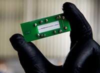
Austin uses electrodes on a printed circuit board and passes the ions over them once. The electrodes detect the charge. To calculate mass, he slows the ions as they go through the system and uses the change in velocity to determine m/z. Measurement precision suffers from not doing a multipass measurement, but Austin is willing to pay that price to be able to detect the particles.
The development with the greatest chance of bringing CDMS to more labs involves the use of Thermo Fisher Scientific’s Orbitrap mass analyzer. Scientists published two reports the same month this year on the preprint server bioRxiv showing that a version of CDMS can be done with Thermo Fisher’s analyzer (bioRxiv 2019, DOI: 10.1101/715425 and DOI: 10.1101/717413). The former was published by Michael Senko and others of Thermo Fisher Scientific and Neil Kelleher’s group at Northwestern University. The latter was published by a team led by Albert Heck of Utrecht University. Kelleher’s interest in CDMS lies in being able to resolve large protein complexes and proteoforms, specific molecular forms of intact proteins. A given protein can have multiple proteoforms, such as different posttranslational modifications.
Complex mixtures of proteoforms push the limits of conventional mass spectrometry. For instance, Kelleher and his team use so-called top-down proteomics to study proteoforms. This means that the researchers analyze intact proteins instead of cutting them into smaller peptides, as is typically done in proteomics to make samples more manageable. Chopping proteins up can result in the loss of the modifications Kelleher wants to study.
Doing top-down proteomics with conventional mass spectrometry, though, yields a thicket of unresolvable peaks in m/z spectra that Kelleher refers to as “the hump of death.” His team can’t extract data from such spectra. CDMS could provide the resolution needed to make sense of proteoform mixtures.
The changes needed to do CDMS on an Orbitrap turn out to be relatively simple ones—letting in fewer ions and lowering the pressure of the trap. The mass analyzer itself doesn’t need to change, because normal Orbitrap operation involves detecting the charges of ions in the trap. But that detection is of large packets of ions at a similar m/z. CDMS requires detecting the charges of individual ions.“We drop the pressure much lower than we normally would because we want to make sure that we can observe an ion for a long time” before it bumps into another ion and potentially changes the reading, Senko says. To let fewer ions into the trap, the scientists either dilute the sample or open the trap entrance for microseconds instead of the usual milliseconds, Kelleher says. They let 120 ions on average into the Orbitrap at a time.
The Thermo Fisher and Northwestern team’s method can assign the charge states of highly charged ions and do it correctly more than 96% of the time. For each instrument, the researchers establish a calibration curve by measuring electrical signals for ions with known charge states and then use the calibration curve to determine unknown charge states for ions in a sample.
Heck’s team at Utrecht University has also used an Orbitrap for CDMS. The researchers similarly establish a charge-state calibration curve but determine that curve and unknown charge states in a slightly different way. They used their calibration to resolve complex mixtures of immunoglobulin G (IgG) and IgM oligomers, ribosome particles, and empty and genome-packed virus assemblies. The ability to do CDMS on a commercial instrument has the possibility of bringing the method to labs that might not want to build their own instruments. But don’t expect a new instrument immediately. “It’s not on our road map as a product right now,” Thermo Fisher’s Senko says. “It’s still very much in our research pipeline.” For those who are impatient, Heck says, “We will make all the software we developed to perform CDMS on an Orbitrap available, which means that all labs having an Orbitrap with extended mass range would be able to do it.”


Join the conversation
Contact the reporter
Submit a Letter to the Editor for publication
Engage with us on Twitter