Advertisement
Grab your lab coat. Let's get started
Welcome!
Welcome!
Create an account below to get 6 C&EN articles per month, receive newsletters and more - all free.
It seems this is your first time logging in online. Please enter the following information to continue.
As an ACS member you automatically get access to this site. All we need is few more details to create your reading experience.
Not you? Sign in with a different account.
Not you? Sign in with a different account.
ERROR 1
ERROR 1
ERROR 2
ERROR 2
ERROR 2
ERROR 2
ERROR 2
Password and Confirm password must match.
If you have an ACS member number, please enter it here so we can link this account to your membership. (optional)
ERROR 2
ACS values your privacy. By submitting your information, you are gaining access to C&EN and subscribing to our weekly newsletter. We use the information you provide to make your reading experience better, and we will never sell your data to third party members.
Biological Chemistry
Fluorescent Dye Helps Visualize Extracellular Matter
June 9, 2008
| A version of this story appeared in
Volume 86, Issue 23

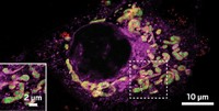
Until now, there hasn’t been a general fluorescent dye that scientists could use to stain the material secreted by cells and view it under a confocal microscope. This extracellular material plays a key role in the fate of cells, including stem cell differentiation and cancer progression. Current dyeing methods, including antibody-targeted staining, have been limited to visualizing one component or only certain kinds of tissues. But now a research team led by Jie Shen at Johannes Gutenberg University and Klaus Müllen at Max Planck Institute for Polymer Research, both in Mainz, Germany, has solved this problem by preparing a positively charged macromolecule that fluoresces red when its amine groups bond to negatively charged extracellular material (J. Am. Chem. Soc., DOI: 10.1021/ja8022362). The water-soluble dye is composed of a central perylene chromophore, a polyphenylene dendrimer scaffold, and a polymer shell bearing multiple amine groups. The researchers used the dye to stain fruit fly tissue as well as bovine collagen, finding that the dye is compatible with antibody-based staining. They suggest that their dye could be useful in multiple-channel fluorescence imaging, and they plan to create different analogs for specific biological applications.


Join the conversation
Contact the reporter
Submit a Letter to the Editor for publication
Engage with us on Twitter